nanotechnology

'Nanocantilevers' yield surprises critical for designing new detectors

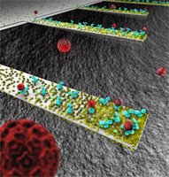
This rendition depicts an array of tiny, diving-boardlike devices called nanocantilevers. The devices are coated with antibodies to capture viruses, which are represented as red spheres. New findings about the behavior of the cantilevers could be crucial in designing a new class of ultra-small sensors for detecting viruses, bacteria and other pathogens. (Image generated by Seyet, LLC)This rendition depicts an array of tiny, diving-boardlike devices called nanocantilevers. The devices are coated with antibodies to capture viruses, which are represented as red spheres.
New findings about the behavior of the cantilevers could be crucial in designing a new class of ultra-small sensors for detecting viruses, bacteria and other pathogens. (Image generated by Seyet, LLC)
Nanocantilevers High Resolution Image.

WEST LAFAYETTE, Ind. — Researchers at Purdue University have made a discovery about the behavior of tiny structures called nanocantilevers that could be crucial in designing a new class of ultra-small sensors for detecting viruses, bacteria and other pathogens.

The nanocantilevers, which resemble tiny diving boards made of silicon, could be used in future detectors because they vibrate at different frequencies when contaminants stick to them, revealing the presence of dangerous substances. Because of the nanocantilever's minute size, it is more sensitive than larger devices, promising the development of advanced sensors that detect minute quantities of a contaminant to provide an early warning that a dangerous pathogen is present

The researchers were surprised to learn that the cantilevers, coated with antibodies to detect certain viruses, attract different densities — or quantity of antibodies per area — depending on the size of the cantilever. The devices are immersed into a liquid containing the antibodies to allow the proteins to stick to the cantilever surface.

"But instead of simply attracting more antibodies because they are longer, the longer cantilevers also contained a greater density of antibodies, which was very unexpected," said Rashid Bashir, a researcher at the Birck Nanotechnology Center and a professor of electrical and computer engineering and biomedical engineering at Purdue University. The research also shows that the density is greater toward the free end of the cantilevers.

The engineers found that the cantilevers vibrate faster after the antibody attachment if the devices have about the same nanometer-range thickness as the protein layer. Moreover, the longer the protein-coated nanocantilever, the faster the vibration, which could only be explained if the density of antibodies were to increase with increasing lengths, Bashir said. The research group also proved this hypothesis using optical measurements and then worked with Ashraf Alam, a researcher at the Birck Nanotechnology Center and professor of electrical and computer engineering, to develop a mathematical model that describes the behavior.

The information will be essential to properly design future "nanomechanical" sensors that use cantilevers, Bashir said.

Findings are detailed in a research paper appearing online today (Monday, Aug. 28) in Proceedings of the National Academy of Sciences. The paper was authored by Amit K. Gupta, a former Purdue doctoral student working with Bashir and now a postdoctoral researcher at Harvard University; Pradeep R. Nair, a doctoral student in electrical and computer engineering; Demir Akin, research assistant professor of biomedical engineering; Michael Ladisch, Distinguished Professor of Agricultural and Biological Engineering with a joint appointment in the Weldon School of Biomedical Engineering; Steven Broyles, a professor of biochemistry; Alam and Bashir.

The work, funded by the National Institutes of Health, is aimed at developing advanced sensors capable of detecting minute quantities of viruses, bacteria and other contaminants in air and fluids by coating the cantilevers with proteins, including antibodies that attract the contaminants. Such sensors will have applications in areas including environmental-health monitoring in hospitals and homeland security. So-called "lab-on-a-chip" technologies could make it possible to replace bulky lab equipment with miniature sensors, saving time, energy and materials. Thousands of the cantilevers can be fabricated on a 1-square-centimeter chip, Bashir said.

The cantilevers studied in the recent work range in length from a few microns to tens of microns, or millionths of a meter, and are about 20 nanometers thick, which is also roughly the thickness of the antibody coating. A nanometer is a billionth of a meter, or approximately the length of 10 hydrogen atoms strung together.

A cantilever naturally "resonates," or vibrates at a specific frequency, depending on its mass and mechanical properties. The mass changes when contaminants land on the devices, causing them to vibrate at a different "resonant frequency, " which can be quickly detected. Because certain proteins attract only specific contaminants, the change in vibration frequency means a particular contaminant is present.

Ordinarily, when using cantilevers that are on a thickness scale of microns or larger, attaching mass causes the resonant frequency to decrease, which is the opposite of what occurs with the nanoscale-thickness cantilevers. Researchers believe the unexpected behavior is a result of the antibodies being about the same thickness as the ultra-thin nanocantilevers, meaning their vibration is more profoundly affected than a more massive cantilever would be by the attachment of the antibodies.

"The conclusion is that when the attached mass is as thick as the cantilever, then you not only affect the mass but you also affect a key property called the net stiffness constant and the resonant frequency can actually go up," Bashir said.

Gupta measured the cantilever's vibration frequency using an instrument called a laser Doppler vibrometer, which detects changes in the cantilever's velocity as it vibrates. The researchers then treated the antibodies with a fluorescent dye and took images of the proteins on the cantilever's surface, proving that the density increases with longer cantilevers

Nair and Alam then developed a mathematical model to explain why the density increases as the area of the cantilever rises. The model uses a "diffusion reaction equation" to simulate the antibodies sticking to the cantilever's surface.

The research is based at the Birck Nanotechnology Center at Discovery Park, the university's hub for interdisciplinary research.

Writer Emil Venere, (765) 494-4709, venere@purdue.edu
Sources: Rashid Bashir, (765) 496-6229, bashir@ecn.purdue.edu
Amit Gupta, (765) 404-5141, agupta@ecn.purdue.edu
Ashraf Alam, (765) 494-5988, alam@purdue.edu
Purdue News Service: (765) 494-2096; purduenews@purdue.edu
nano GALLERY

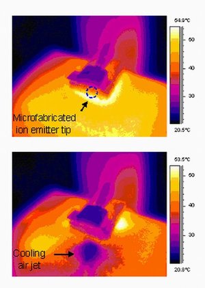
Infrared images show how a new UW micro-pump cools a heated surface: (Top) The air pump is off. (Bottom) The air pump is on.University of Washington, Infrared images show how a new UW micro-pump cools a heated surface: (Top) The air pump is off. (Bottom) The air pump is on. highest resolution version of this photo (print ready).
Tiny ion pump sets new standard in cooling hot computer chips FULL TEXT, ion pump cooling hot computer chips.

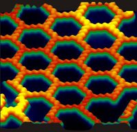
Caption: Anthraquinone molecules form chains of molecules that weave themselves into a sheet of hexagons on a polished copper surface. Credit: Ludwig Bartels's research group, UCR, Usage Restrictions: None.Caption: Anthraquinone molecules form chains of molecules that weave themselves into a sheet of hexagons on a polished copper surface. Credit: Ludwig Bartels's research group, UCR, Usage Restrictions: None.
FULL TEXT, Honeycomb Network Comprised of Anthraquinone Molecules, Molecules spontaneously form honeycomb network featuring pores of unprecedented size

This rendition depicts an array of tiny, diving-boardlike devices called nanocantilevers. The devices are coated with antibodies to capture viruses, which are represented as red spheres. New findings about the behavior of the cantilevers could be crucial in designing a new class of ultra-small sensors for detecting viruses, bacteria and other pathogens. (Image generated by Seyet, LLC)This rendition depicts an array of tiny, diving-boardlike devices called nanocantilevers. The devices are coated with antibodies to capture viruses, which are represented as red spheres.
New findings about the behavior of the cantilevers could be crucial in designing a new class of ultra-small sensors for detecting viruses, bacteria and other pathogens. (Image generated by Seyet, LLC)
Nanocantilevers High Resolution Image. FULL TEXT 'Nanocantilevers' yield surprises critical for designing new detectors.

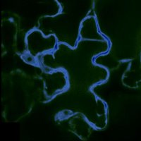
This false-color image shows a cell from the epidermis of an Arabidopsis thaliana plant; the cell has been marked with fluorescent imaging sensors designed to detect the sugar glucose. In this image, only the densely packed interior of the cell in which most metabolic functions occur—called the cytosol—is targeted by the glucose sensors. The dark area sits inside the vacuole—a large storage organelle that can occupy up to 90% of the cell’s volume. (Image courtesy Sylvie Lalonde and Wolf Frommer;)This false-color image shows a cell from the epidermis of an Arabidopsis thaliana plant; the cell has been marked with fluorescent imaging sensors designed to detect the sugar glucose.
In this image, only the densely packed interior of the cell in which most metabolic functions occur—called the cytosol—is targeted by the glucose sensors. The dark area sits inside the vacuole—a large storage organelle that can occupy up to 90% of the cell’s volume. (Image courtesy Sylvie Lalonde and Wolf Frommer; click for higher resolution.) FULL TEXT Sugar metabolism tracked in living plant tissues, in real time.

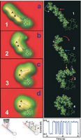
Scientists used a scanning tunneling microscope to manipulate chlorophyll-a into four positions. art by: Saw-Wai HlaNanoscientists Create Biological Switch from Spinach Molecule, Scientists used a scanning tunneling microscope to manipulate chlorophyll-a into four positions. art by: Saw-Wai Hla, Tuesday Sep 05, 2006, by Andrea Gibson. FULL TEXT Nanoscientists Create Biological Switch from Spinach Molecule

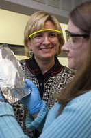
Jamie Mullally '07, right, a Cornell Presidential Research Scholar, and Margaret Frey, assistant professor of textiles and apparel, examine a nonwoven nanofiber fabric on aluminum foil backing. Mullally will complete an honors thesis on the biorecognition fabrics in spring '07. Copyright © Cornell UniversityJamie Mullally '07, right, a Cornell Presidential Research Scholar, and Margaret Frey, assistant professor of textiles and apparel, examine a nonwoven nanofiber fabric on aluminum foil backing. Mullally will complete an honors thesis on the biorecognition fabrics in spring '07. Copyright © Cornell University. FULL TEXT, Biodegradable napkin, featuring nanofibers, may detect biohazards

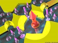
Nanoscale metallic electrodes (in yellow) can be used to confine electrons in small regions, forming quantum dots. Two quantum dots connected to each other form a double quantum dot. In this case, one of the dots is in the Kondo state, in which the magnetic moment of the confined electron (large red arrow) is compensated ('screened') by the magnetic moment of surrounding electrons, resulting in a zero net magnetic moment for the entire system. art by: Luis Dias/Ohio UniversityNanoscale metallic electrodes (in yellow) can be used to confine electrons in small regions, forming quantum dots. Two quantum dots connected to each other form a double quantum dot.
In this case, one of the dots is in the Kondo state, in which the magnetic moment of the confined electron (large red arrow) is compensated (“screened”) by the magnetic moment of surrounding electrons, resulting in a zero net magnetic moment for the entire system. art by: Luis Dias/Ohio University. FULL TEXT, Double Quantum Dots Control Kondo Effect

MIT researchers have discovered that certain molecules can attach themselves to metallic carbon nanotubes without interfering with the nanotubes' exceptional ability to conduct electricity. At left, the high conductance state has two molecular orbitals, shown in green. Some molecules even let the nanotube switch between highly conductive, left, and poorly conductive (right, with one red molecular orbital), creating the potential for new applications. Image courtesy / Marzari Lab.Based on a new theory, MIT scientists may be able to manipulate carbon nanotubes --
one of the strongest known materials and one of the trickiest to work with -- without destroying their extraordinary electrical properties. FULL TEXT, scientists tame tricky carbon nanotubes

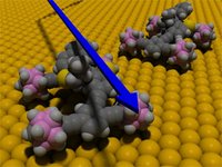
Title: Motorized Nanocar, Credit: Yasuhiro Shirai/Rice UniversityThis animation depicts two motorized nanocars on a gold surface. The nanocar consists of a rigid chassis and four alkyne axles that spin freely
and swivel independently of one another. The wheels are spherical molecules of carbon, hydrogen and boron called p-carborane. FULL TEXT, Nanocar inventor named top nanotech innovator

Caption: In NIST's Einstein-de Haas experiment, the movements of a cantilever were measured with an optical-fiber laser interferometer. The optical fiber is 125 micrometers in diameter, and the end is positioned less than 10 micrometers from the cantilever surface. Credit: Credit: John Moreland/NIST, Usage Restrictions: None.Caption: In NIST's Einstein-de Haas experiment.
the movements of a cantilever were measured with an optical-fiber laser interferometer, The optical fiber is 125 micrometers in diameter, and the end is positioned less than 10 micrometers from the cantilever surface. Credit: Credit: John Moreland/NIST, Usage Restrictions: None. FULL TEXT, Einstein's magnetic effect is measured on microscale

Honeycomb Network Comprised of Anthraquinone Molecules

Molecules spontaneously form honeycomb network featuring pores of unprecedented size

UCR discovery of molecular self-assembly could help develop templates for growing complex structures on surfaces; improve paints and lubricants.

Caption: Anthraquinone molecules form chains of molecules that weave themselves into a sheet of hexagons on a polished copper surface. Credit: Ludwig Bartels's research group, UCR, Usage Restrictions: None.Caption: Anthraquinone molecules form chains of molecules that weave themselves into a sheet of hexagons on a polished copper surface. Credit: Ludwig Bartels's research group, UCR, Usage Restrictions: None.
Riverside, Calif. -- UC Riverside researchers have discovered a new way in which nature creates complex patterns: the assembly of molecules with no guidance from an outside source. Potential applications of the finding are paints, lubricants, medical implants, and processes where surface-patterning at the scale of molecules is desired.

Spreading anthraquinone, a common and inexpensive chemical, on to a flat copper surface, Greg Pawin, a chemistry graduate student working in the laboratory of Ludwig Bartels, associate professor of chemistry, observed the spontaneous formation of a two-dimensional honeycomb network comprised of anthraquinone molecules.

The finding, reported in the Aug. 18 issue of Science, describes a new mechanism by which complex patterns are generated at the nanoscale – 0.1 to 100 nanometers in size, a nanometer being a billionth of a meter – without any need for expensive processes such as lithography.

"We know that some of the most striking phenomena in nature, like the colors on a butterfly wing, come about by the regular arrangement of atoms and molecules," said Pawin, the first author of the paper. "But what physical and chemical processes guide their arrangement? Anthraquinone showed us how such patterns can form easily and spontaneously."

Over a span of several years, Bartels's research group tested a multitude of molecules for pattern formation at the nanoscale. The group found that, generally, these molecules tended to become lumps, forming uninteresting islands of molecules lying side by side.

Anthraquinone molecules, however, form chains that weave themselves into a sheet of hexagons on the copper surface, forming a network similar to chicken wire. The precise shape of the network is governed by a delicate balance between forces of attraction and repulsion operating on the molecules.

"The honeycomb pattern that the anthraquinone molecules produce is open, meaning it has big pores, or cavities, enclosed by the hexagonal rings," Pawin said. "Such patterns have never been observed before. Rather, the common belief was that they cannot be generated. But anthraquinone shows that we can use chemistry to engineer molecules that self-assemble into structures with pores that are many times larger than the individual molecules themselves. With judicious engineering of the relation between the strength of the attraction and repulsion, we could tailor film patterns and pore sizes almost at will."

Patterning of surfaces is important for many applications. The friction that water or air experience when flowing over a surface crucially depends on the microscopic structure of the surface. Biological cells and tissue grow easily on surfaces of some patterns while rejecting other patterns and completely flat surfaces.

In their research the UCR chemists first cleaned the copper surface, creating an extremely slippery surface. Then they deposited anthraquinone molecules onto it. Next, the surface with the molecules was annealed to spread the molecules. During cool-down to the temperature of liquid nitrogen, the hexagonal pattern emerged.

Pawin also developed a computer model to understand not only why the anthraquinone molecules lined up in rows that ultimately arranged themselves into a honeycomb network, but also how anthraquinone molecules are prevented from taking up space inside the pores.

"The precise pattern anthraquinone forms depends on a delicate balance between the attraction between the anthraquinone molecules and the substrate-mediated forces that ultimately disperse these molecules," said Bartels, a member of UCR's Center for Nanoscale Science and Engineering. "By fine-tuning this balance, it should be possible to produce a wide variety of patterns of different sizes."

In the future, Pawin and Bartels plan on investigating how chemical modifications of anthraquinone can produce novel patterns. "In addition, we would like to form the hexagonal network at higher temperatures and be able to control the size of the hexagons," Pawin said. "We also want to extend our research to include surfaces other than copper and determine if there are molecules similar to anthraquinone that assemble spontaneously into sheets on them."

Besides Pawin and Bartels, Kin L. Wong and Ki-Young Kwon of Bartels's research group participated in the study, which was supported by a grant from the National Science Foundation. Pawin first started working in Bartels's laboratory in 2000 as an undergraduate. This fall, he will be a second-year graduate student at UCR.

Details of the study

Anthraquinone molecules consist of three fused benzene rings with one oxygen atom on each side. An organic compound, anthraquinone is widely used in the pulp industry for turning cellulose from wood into paper. It is also the parent substance of a large class of dyes and pigments. Its chemical formula is C14H8O2.

The pore diameter of the honeycomb network of anthraquinone molecules is about 50 Å. The attractive interaction between the molecules stems from hydrogen bridge bonding, a phenomenon common in nature and fundamental to all life (e.g., by holding DNA helixes together) but which occurs here in a slightly unconventional and novel form. The substrate-meditated repulsive interactions are: (a) expansion of the copper surface because the anthraquinone molecules 'dig' their oxygen 'heels' into it; (b) electrostatic repulsion due to slightly negative charging of the anthraquinone molecules; or (c) a combination thereof.

The UCR study used a scanning tunneling microscope in Bartels's laboratory which can image individual molecules at great precision. An individual anthraquinone molecule appears as an almost rectangular feature with slightly rounded edges. The sides of each hexagon consist of three parallel anthraquinone molecules. The vertices consist of three anthraquinone molecules that form a triangle. Each hexagon encloses more than 200 atoms of the copper substrate. ###


The University of California, Riverside is a major research institution. Key areas of research include nanotechnology, health science, genomics, environmental studies, digital arts and sustainable growth and development. With a current undergraduate and graduate enrollment of more than 16,600, the campus is projected to grow to 21,000 students by 2010. Located in the heart of Inland Southern California, the nearly 1,200-acre, park-like campus is at the center of the region's economic development. Visit ucr.edu/ or call 951-UCR-NEWS for more information. Media sources are available at mediasources.ucr.edu/.

Contact: Iqbal Pittalwala iqbal@ucr.edu 951-827-6050 University of California - Riverside

Sugar metabolism tracked in living plant tissues, in real time

This false-color image shows a cell from the epidermis of an Arabidopsis thaliana plant; the cell has been marked with fluorescent imaging sensors designed to detect the sugar glucose. In this image, only the densely packed interior of the cell in which most metabolic functions occur—called the cytosol—is targeted by the glucose sensors. The dark area sits inside the vacuole—a large storage organelle that can occupy up to 90% of the cell’s volume. (Image courtesy Sylvie Lalonde and Wolf Frommer;)This false-color image shows a cell from the epidermis of an Arabidopsis thaliana plant; the cell has been marked with fluorescent imaging sensors designed to detect the sugar glucose.
In this image, only the densely packed interior of the cell in which most metabolic functions occur—called the cytosol—is targeted by the glucose sensors. The dark area sits inside the vacuole—a large storage organelle that can occupy up to 90% of the cell’s volume. (Image courtesy Sylvie Lalonde and Wolf Frommer; click for higher resolution.)

Stanford, CA – Scientists at Carnegie’s Department of Plant Biology have made the first real-time observations of sugars in the cells of intact and living plant tissues. With the help of groundbreaking imaging techniques, the group has determined that plants maintain extremely low levels of sugar in their roots—as much as 100,000 times lower than previous estimates. The new technology will enable new studies of sugar metabolism in plants, which will inform the effort to engineer higher crop yields for food and biofuel production.

Led by Carnegie staff member Wolf Frommer, the researchers designed genetically-encoded fluorescent tags to monitor glucose, an important sugar, in leaf and root tissues of the model plant Arabidopsis thaliana. The technique has allowed the researchers to track glucose over time and space at unprecedented detail, in living and undisturbed plant tissues. The work appears in the September issue of the journal Plant Cell*. The group has also developed a FRET sensor for sucrose, a major transport sugar in plants. This work will appear in the September issue of the Journal of Biological Chemistry**.

“Until now, we have had few clues regarding how much sugar is in an individual cell in a multicellular plant,” Frommer said. “We normally grind up a leaf or a root and average the information for all cells, but if sugar levels rise in one cell and drop in another, we would see no change in this average.” Also, because the cell can distribute sugar among subcellular organelles, it is nearly impossible to know how much sugar is in any cell compartment at a given time.
“Time resolution is another problem,” Frommer added. “We can sample tissue at intervals, but if the sugar changes in waves, we might miss the right time point. Our new technology addresses all of these problems by measuring sugar flux in real time in individual cells, with subcellular resolution.”

Frommer and his colleagues have used similar imaging tags, called fluorescent resonance energy transfer (FRET) sensors, to track sugars and neurotransmitters in animal cells. Most recently, the group used FRET sensors to study glutamate, an important mammalian neurotransmitter. Frommer has tracked glucose in cultured mammalian cells, but until now, plant tissues had proven problematic because of interference from the plants’ virus defense mechanisms, as well as high background fluorescence in some plants.

To surmount these issues, Frommer’s team dramatically improved the sensors, while inserting them in mutant Arabidopsis plants with disabled defense genes. The fluorescent tags worked well where they had failed before.

“It may not be ideal to use defense-mutant plants—the ideal would be for the sensors to work in any wild-type genetic background,” Frommer explained. “But proving that the sensors can work in plants is an important first step. Now we can begin addressing important questions about the way plants manage sugar distribution while we continue to improve the sensors.”

In preliminary experiments, Frommer’s group compared fluctuations in glucose levels in root tissue and leaf epidermis—the topmost layer that absorbs sunlight—and found that the plant maintained glucose at higher levels in leaf tissue than in roots. In fact, the researchers found that root cells contain sugar at concentrations at least 100,000 times lower than previous estimates.

FRET sensors are encoded by genes that, in theory, can be engineered into any cell line or organism. They are made of two fluorescent proteins that produce different colors of light—one cyan and one yellow—connected by a third protein that resembles a hinged clam shell. The two fluorescent proteins are derived from jellyfish, and the third from a bacterium; the shape of the clam shell protein determines which sugar or other molecule the sensor can detect. When a target molecule such as glucose or sucrose binds to the third protein, the hinge opens, changing the distance and orientation of the fluorescent proteins. This physical change affects the energy transfer between the cyan and yellow markers.

When the researchers hit the tags with light of a specific wavelength, the cyan tag starts to fluoresce. If the yellow tag is close enough, the cyan tag will transfer its energy to the yellow tag, causing it to resonate and fluoresce as well. This energy transfer affects how much cyan and yellow fluorescence can be seen, and by calculating this ratio, researchers can accurately track molecules such as glucose and sucrose in both time and space.

“The strength of this technology lies in its elegant simplicity; with the power of computational design, we can potentially design FRET tags to detect virtually any small molecule in living cells,” Frommer said. “Imaging techniques like this are the next frontier in the study of metabolism, and will help to answer some of the most pressing questions on plant biologists’ minds, such as the role of individual genes in the distribution of sugars. This in turn can help us engineer plants to produce more biomass.”

Carnegie Contact: Dr. Wolf Frommer; wfrommer@stanford.edu or (650) 325-1521 x208, News ReleaseThursday, August 31, 2006

*For a copy of the Plant Cell paper, go to: plantcell.org/cgi/content/short/

**For a copy of the Journal of Biological Chemistry paper, go to: jbc.org/cgi/reprint/

The two projects were supported by grants from the U.S. Department of Energy, the National Institutes of Health, and the European Science award of the Körber Foundation, Hamburg.

The Carnegie Institution of Washington (carnegieinstitution.org) has been a pioneering force in basic scientific research since 1902. It is a private, nonprofit organization with six research departments throughout the U.S. Carnegie scientists are leaders in plant biology, developmental biology, astronomy, materials science, global ecology, and Earth and planetary science. The Department of Plant Biology is located at 260 Panama St., Stanford, CA 94305

Nanoscientists Create Biological Switch from Spinach Molecule

Scientists used a scanning tunneling microscope to manipulate chlorophyll-a into four positions. art by: Saw-Wai HlaNanoscientists Create Biological Switch from Spinach Molecule, Scientists used a scanning tunneling microscope to manipulate chlorophyll-a into four positions. art by: Saw-Wai Hla, Tuesday Sep 05, 2006, by Andrea Gibson
ATHENS, Ohio – Nanoscientists have transformed a molecule of chlorophyll-a from spinach into a complex biological switch that has possible future applications for green energy, technology and medicine.

The study offers the first detailed image of chloropyhll-a – the main ingredient in the photosynthesis process – and shows how scientists can use new technology to manipulate the configuration of the spinach molecule in four different arrangements, report Ohio University physicists Saw-Wai Hla and Violeta Iancu in today’s early edition of the journal Proceedings of the National Academy of Sciences.

The scientists used a scanning tunneling microscope to image chlorophyll-a and then injected it with a single electron to manipulate the molecule into four positions, ranging from straight to curved, at varying speeds. (View a movie here) Though the Ohio University team and others have created two-step molecule switches using scanning tunneling microscope manipulation in the past, the new experiment yields a more complex multi-step switch on the largest organic molecule to date.

The work has immediate implications for basic science research, as the configuration of molecules and proteins impacts biological functions. The study also suggests a novel route for creating nanoscale logic circuits or mechanical switches for future medical, computer technology or green energy applications, said Hla, an associate professor of physics.

“It’s important to understand something about the chlorophyll-a molecule for origin of life and solar energy conversion issues,” he said.

The study was funded by Ohio University’s Nanobiotechnology Initiative and the U.S. Department of Energy. Hla is a member of the university’s Quantitative Biology Institute and Nanoscale & Quantum Phenomena Institute. Iancu is a doctoral candidate in the Department of Physics and Astronomy.

Additional information, Contact: Saw-Wai Hla, (740) 593-1727, hla@ohio.edu

Related News:

Biodegradable napkin, featuring nanofibers, may detect biohazards

CU biodegradable wipe would quickly detect biohazards, from avian flu to E. coli, By Susan Lang

Jamie Mullally '07, right, a Cornell Presidential Research Scholar, and Margaret Frey, assistant professor of textiles and apparel, examine a nonwoven nanofiber fabric on aluminum foil backing. Mullally will complete an honors thesis on the biorecognition fabrics in spring '07. Copyright © Cornell UniversityJamie Mullally '07, right, a Cornell Presidential Research Scholar, and Margaret Frey, assistant professor of textiles and apparel, examine a nonwoven nanofiber fabric on aluminum foil backing.
Mullally will complete an honors thesis on the biorecognition fabrics in spring '07. Copyright © Cornell University.

Detecting bacteria, viruses and other dangerous substances in hospitals, airplanes and other commonly contaminated places could soon be as easy as wiping a napkin or paper towel across a surface

"It's very inexpensive, it wouldn't require that someone be highly trained to use it, and it could be activated for whatever you want to find," said Margaret Frey, the Lois and Mel Tukman Assistant Professor of Fiber Science and Apparel Design at Cornell University. "So if you're working in a meat-packing plant, for instance, you could swipe it across some hamburger and quickly and easily detect E. coli bacteria." She reported on the research Sept. 11 at the American Chemical Society's national meeting.

Once fully developed, the biodegradable absorbent wipe would contain nanofibers containing antibodies to numerous biohazards and chemicals and would signal by changing color or through another effect when the antibodies attached to their targets. Users would simply wipe the napkin across a surface; if a biohazard were detected, the surface could be disinfected and retested with another napkin to be sure it was no longer contaminated.

In work conducted with Yong Joo, assistant professor of chemical and biomolecular engineering, and Antje Baeumner, associate professor of biological and environmental engineering, both at Cornell, Frey developed nanofibers with platforms made of biotin, a part of the B vitamin complex, and the protein streptavidin, which can hold the antibodies. Composed of a polymer compound made from corn, the nanofibers could be incorporated into conventional paper products to keep costs low. Nanofibers, with diameters near 100 nanometers (a nanometer is one-billionth of a meter, or about three times the diameter of an atom), provide extremely large surface areas for sensing and increased absorbency compared with conventional fibers.

"The fabric basically acts as a sponge that you can use to dip in a liquid or wipe across a surface," Frey said. "The fabric itself will transport and concentrate the targeted biohazard. As you do that, antibodies in the fabric are going to selectively latch onto whatever pathogen that they match. Using this method we should, in theory, be able to quickly activate the fabric to detect whatever is the hazard of the week, whether it is bird flu, mad cow disease or anthrax."

Frey and her colleagues are still working on ways, such as a color change, for the fabric to signal that it has identified the contaminant.

"We're probably still a few years away from having this ready for the real world," Frey said, "but I really believe there is a place for this type of product that can be used by people with limited training to provide a fast indication of whether a biohazard is present."

This research was supported by the National Research Initiative of the U.S. Department of Agriculture's Cooperative State Research, Education and Extension Service.

### Contact: Blaine Friedlander bpf2@cornell.edu 607-254-8093 Cornell University News Service

Double Quantum Dots Control Kondo Effect

Nano News: Double Quantum Dots Control Kondo Effect, Thursday Sep 14, 2006 by ANDREA GIBSON

Nanoscale metallic electrodes (in yellow) can be used to confine electrons in small regions, forming quantum dots. Two quantum dots connected to each other form a double quantum dot. In this case, one of the dots is in the Kondo state, in which the magnetic moment of the confined electron (large red arrow) is compensated ('screened') by the magnetic moment of surrounding electrons, resulting in a zero net magnetic moment for the entire system. art by: Luis Dias/Ohio UniversityNanoscale metallic electrodes (in yellow) can be used to confine electrons in small regions, forming quantum dots. Two quantum dots connected to each other form a double quantum dot.
In this case, one of the dots is in the Kondo state, in which the magnetic moment of the confined electron (large red arrow) is compensated (“screened”) by the magnetic moment of surrounding electrons, resulting in a zero net magnetic moment for the entire system. art by: Luis Dias/Ohio University

ATHENS, Ohio — Two quantum dots connected by wires could help scientists better control the Kondo effect in experiments, according to a study by Ohio University and University of Florida physicists published in a recent issue of Physical Review Letters.

The Kondo effect occurs when electrons become trapped around the magnetic impurities in semiconductor materials, which prompts the electrons to change their spin. This phenomenon has intrigued scientists, as electronic correlations can create interesting and complex behavior in materials.

In the new work, scientists demonstrate how the two quantum dot system can behave in two different and interesting ways: As a simile for a Kondo-effect system where one quantum dot is used to "filter" the effect of the current leads, and as a way to study "pseudo-gapped" systems and correlations in them, which can help scientists understand structures such as superconductors.

“This last part is of great current interest to theorists and experimentalists who are exploring what are called quantum phase transitions, which are changes in systems that alter their behavior dramatically as a function of some parameter while remaining at zero (or very low) temperature,” said Sergio Ulloa, a professor of physics and astronomy at Ohio University.

The study, funded by the National Science Foundation, was conducted by Luis Dias da Silva, Nancy Sandler and Ulloa, all members of the Ohio University’s Nanoscale and Quantum Phenomena Institute, and Kevin Ingersent of the University of Florida.

scientists tame tricky carbon nanotubes

MIT materials scientists tame tricky carbon nanotubes, Deborah Halber, News Office Correspondent, September 15, 2006

MIT researchers have discovered that certain molecules can attach themselves to metallic carbon nanotubes without interfering with the nanotubes' exceptional ability to conduct electricity. At left, the high conductance state has two molecular orbitals, shown in green. Some molecules even let the nanotube switch between highly conductive, left, and poorly conductive (right, with one red molecular orbital), creating the potential for new applications. Image courtesy / Marzari Lab.Based on a new theory, MIT scientists may be able to manipulate carbon nanotubes --
one of the strongest known materials and one of the trickiest to work with -- without destroying their extraordinary electrical properties.

The work is reported in the Sept. 15 issue of Physical Review Letters, the journal of the American Physical Society.

Carbon nanotubes -- cylindrical carbon molecules 50,000 times thinner than a human hair -- have properties that make them potentially useful in nanotechnology, electronics, optics and reinforcing composite materials. With an internal bonding structure rivaling that of another well-known form of carbon, diamonds, carbon nanotubes are extraordinarily strong and can be highly efficient electrical conductors.

The problem is working with them. There is no reliable way to arrange the tubes into a circuit, partly because growing them can result in a randomly oriented mess resembling a bowl of spaghetti.

Researchers have attached to the side walls of the tiny tubes chemical molecules that work as "handles" that allow the tubes to be assembled and manipulated. But these molecular bonds also change the tubes' structure and destroy their conductivity.

Now Young-Su Lee, an MIT graduate student in materials science and engineering, and Nicola Marzari, an associate professor in the same department, have identified a class of chemical molecules that preserve the metallic properties of carbon nanotubes and their near-perfect ability to conduct electricity with little resistance.

Using these molecules as handles, Marzari and Lee said, could overcome fabrication problems and lend the nanotubes new properties for a host of potential applications as detectors, sensors or components in novel optoelectronics.

Playing with atoms
Marzari and Lee use the fundamental laws of quantum mechanics to simulate material properties that are difficult or impossible to measure, such as molten lava in the Earth's core or atomic motion in a fast chemical reaction. Then they run these simulations on interconnected PCs and use the results to optimize and engineer novel materials such as electrodes for fuel cells and polymers that contract and expand like human muscles.

With the help of a powerful algorithm created by Lee and published last year in Physical Review Letters, the theorists focused on solving some of the problems of working with carbon nanotubes.

Like fuzzy balls and Velcro, the hexagon of carbon that makes up a nanotube has a predilection for clinging to other hexagons. One of the many challenges of working with the infinitesimally small tubes is that they tend to stick to each other.

Attaching a molecule to the sidewall of the tube serves a double purpose: It stops nanotubes from sticking so they can be processed and manipulated more easily, and it allows researchers to control and change the tubes' electronic properties. Still, most such molecules also destroy the tubes' conductance because they make the tube structurally more similar to a diamond, which is an insulator, rather than to graphite, a semi-metal.

Lee and Marzari used Lee's algorithm to identify a class of "molecular handles" (carbenes and nitrenes) that stop this from happening and preserve the tubes' original conductivity. "We now have a way to attach molecules that allows us to manipulate the nanotubes without losing their conductance," Marzari said.

Carbenes and nitrenes work by breaking a molecular bond on the nanotube's wall while creating their own new bond to the tube. This process -- one bond formed, one bond lost -- restores the perfect number of bonds each carbon atom had in the original tube and "conductance is recovered," Marzari said.

Some molecular handles can even transform between a bond-broken and a bond-intact state, allowing the nanotubes to act like switches that can be turned on or off in the presence of certain substances or with a laser beam. "This direct control of conductance may lead to novel strategies for the manipulation and assembly of nanotubes in metallic interconnects, or to sensing or imaging devices that respond in real-time to optical or chemical stimuli," Marzari said.

The next step is for experiments to confirm that the approach works.

This work is supported by the MIT Institute for Soldier Nanotechnologies and the National Science Foundation

Einstein's magnetic effect is measured on microscale

Caption: In NIST's Einstein-de Haas experiment, the movements of a cantilever were measured with an optical-fiber laser interferometer. The optical fiber is 125 micrometers in diameter, and the end is positioned less than 10 micrometers from the cantilever surface. Credit: Credit: John Moreland/NIST, Usage Restrictions: None.Caption: In NIST's Einstein-de Haas experiment.
the movements of a cantilever were measured with an optical-fiber laser interferometer, The optical fiber is 125 micrometers in diameter, and the end is positioned less than 10 micrometers from the cantilever surface. Credit: Credit: John Moreland/NIST, Usage Restrictions: None.

A gyromagnetic effect discovered by Albert Einstein and Dutch physicist Wander Johannes de Haas--the rotation of an object caused by a change in magnetization--has been measured at micrometer-scale dimensions for the first time at the National Institute of Standards and Technology (NIST). The new method may be useful in the development and optimization of thin film materials for read heads, memories and recording media for magnetic data storage and spintronics, an emerging technology that relies on the spin of electrons instead of their charge as in conventional electronics.

The Einstein-de Haas effect was first observed in experiments reported in 1915, in which a large iron cylinder suspended by a glass wire was made to rotate by an alternating magnetic field applied along the cylinder's central axis. By contrast, the NIST experiments, described in the Sept. 18 issue of Applied Physics Letters,* measured the Einstein-de Haas effect in a ferromagnetic thin film only 50 nanometers thick deposited on a microcantilever--a tiny beam anchored at one end and projecting into the air. An alternating magnetic field induced changes in the magnetic state of the thin film, and the resulting torque bent the cantilever up and down by just a few nanometers.

Using a laser interferometer to measure the movements of the cantilever and comparing those data to changes in the magnetic state of the material, researchers were able to determine the "magnetomechanical ratio," or the extent to which the material twists in response to changes in its magnetic state. The magnetomechanical ratio is related to another important parameter, the "g-factor," a measure of the internal magnetic rotation of the electrons in a material in a magnetic field.

The magnetomechanical ratio and the g-factor are critical in understanding magnetization dynamics and designing magnetic materials for data storage and spintronics applications, but they are extremely difficult to determine accurately because of many potential complicating effects. The NIST experiments provide a proof-of-concept for using the Einstein-de Haas effect to determine the magnetomechanical ratio and the related g-factor in thin ferromagnetic films. The researchers note that a number of improvements are possible, such as operating the cantilever system in a vacuum to reduce the effects of any changes in temperature. ###

* T.M. Wallis, J. Moreland and P. Kabos. 2006. Einstein-de Haas effect in a NiFe film deposited on a microcantilever. Applied Physics Letters. Sept. 18.

Related Posts Plugin for WordPress, Blogger...

Visitors


free counter
Advertise my site free UseAds.com! Add & submit url & exchange text links + increase traffic & improve page rank!

Advertising my web site free online UseAds.com - Add & submit url & exchange text links + increase traffic & promotion marketing website